Some Of You May Be Familiar With Model Organisms In Biology But Even So, You May Think About Mice, Rabbits
Some of you may be familiar with model organisms in biology but even so, you may think about mice, rabbits or flies rather than ctenophores. The whole purpose of having a model organism is to be able to understand particular biological functions/processes by using an organism that can be maintained easily, has a relatively short generation time and has its genome sequenced (this allows us to really understand their genetic makeup). Since this species of ctenophore (Mnemiopsis leidyi) has had its genome sequenced it allows us to identify key genes/proteins and try to determine their function.
The work I am currently doing for my project is focused on understanding the origin of the nervous system.
There's been a long standing debate amongst scientists over which species of animal first diverged from all other metazoans whether it be sponges or ctenophores. For a long time it has been thought that sponges are the sister group to all metazoans, although more recently studies have suggested that ctenophores are. Sponges are really simple animals that lack nervous systems, whereas ctenophores are more complex and have a nervous system. If ctenophores are then in fact found to be the sister group to all other metazoans, it poses the questions about whether the complex structures such as neurons and synapses evolved once or multiple times independently?
If you check you can see a diagram showing what I mean by the "sister group" to all metazoans. The first pic identifies sponges as the sister group, but with more analysis on a molecular basis, the 3rd pic could be possible.
Since most of the studies on neurons and nervous systems more generally are focused on metazoans, the work at this lab uses ctenophores to understand more about their complex biology with the aim of understanding the origins of neurons.


More Posts from Invincibleworld and Others


I am the owner
Of this damage, the weapon
Fired to make the wound
Eyes full of apologies
Belie my gunpowder smile


Highway system or fungal contaminants in an in-clinic diffquik stain?
A great example of branching, septate fungal hyphae and why in-clinic cytology dyes should be changed regularly! Fungi love to grow in them and can easily be misinterpreted as pathological!


Lab lately
Bye jet leg and hello experiments. Back to the lab like I never left. Summer hasn't been summering in the States so I made the most of sitting on the balcony enjoying the sun for 1 day
But l'm not really outside this summer soooo catch me INSIDE
It's still so weird to me that this is what a bacteriophage actually looks

Like, tf do you mean it's not just a diagram, and it really looks like this


Literally on the day of Halloween I have managed to chance upon a cell in a splenic impression smear that looks vaguely like fatso from Casper and I couldn't be happier

Suspicions confirmed: Common cause for brain tumors in children
An overactive signaling pathway is a common cause in cases of pilocytic astrocytoma, the most frequent type of brain cancer in children. This was discovered by a network of scientists coordinated by the German Cancer Research Center (as part of the International Cancer Genome Consortium, ICGC). In all 96 cases studied, the researchers found defects in genes involved in a particular pathway. Hence, drugs can be used to help affected children by blocking components of the signaling cascade. The project is funded by the German Cancer Aid (Deutsche Krebshilfe) and the Federal Ministry of Education and Research (BMBF). The findings are published in the latest issue of the journal “Nature Genetics”.
Brain cancer is the primary cause of cancer mortality in children. Even in cases when the cancer is cured, young patients suffer from the stress of a treatment that can be harmful to the developing brain. In a search for new target structures that would create more gentle treatments, cancer researchers are systematically analyzing all alterations in the genetic material of these tumors. This is the mission of the PedBrain consortium, which was launched in 2010. Led by Professor Stefan Pfister from the German Cancer Research Center (Deutsches Krebsforschungszentrum, DKFZ), the PedBrain researchers have now published the results of the first 96 genome analyses of pilocytic astrocytomas.
Pilocytic astrocytomas are the most common childhood brain tumors. These tumors usually grow very slowly. However, they are often difficult to access by surgery and cannot be completely removed, which means that they can recur. The disease may thus become chronic and have debilitating effects for affected children.
In previous work, teams of researchers led by Professor Dr. Stefan Pfister and Dr. David Jones had already discovered characteristic mutations in a major proportion of pilocytic astrocytomas. All of the changes involved a key cellular signaling pathway known as the MAPK signaling cascade. MAPK is an abbreviation for “mitogen-activated protein kinase.” This signaling pathway comprises a cascade of phosphate group additions (phosphorylation) from one protein to the next – a universal method used by cells to transfer messages to the nucleus. MAPK signaling regulates numerous basic biological processes such as embryonic development and differentiation and the growth and death of cells.
“A couple of years ago, we had already hypothesized that pilocytic astrocytomas generally arise from a defective activation of MAPK signaling,” says David Jones, first author of the publication. “However, in about one fifth of the cases we had not initially discovered these mutations. In a whole-genome analysis of 96 tumors we have now discovered activating defects in three other genes involved in the MAPK signaling pathway that have not previously been described in astrocytoma.“
“Aside from MAPK mutations, we do not find any other frequent mutations that could promote cancer growth in the tumors. This is a very clear indication that overactive MAPK signals are necessary for a pilocytic astrocytoma to develop,” says study director Stefan Pfister. The disease thus is a prototype for rare cancers that are based on defects in a single biological signaling process.
In total, the genomes of pilocytic astrocytomas contain far fewer mutations than are found, for example, in medulloblastomas, a much more malignant pediatric brain tumor. This finding is in accordance with the more benign growth behavior of astrocytomas. The number of mutations increases with the age of the affected individuals.
About one half of pilocytic astrocytomas develop in the cerebellum, the other 50 percent in various other brain regions. Cerebellar astrocytomas are genetically even more homogenous than other cases of the disease: In 48 out of 49 cases that were studied, the researchers found fusions between the BRAF gene, a central component of the MAPK signaling pathway, and various other fusion partners.
“The most important conclusion from our results,” says study director Stefan Pfister, “is that targeted agents for all pilocytic astrocytomas are potentially available to block an overactive MAPK signaling cascade at various points. We might thus in the future be able to also help children whose tumors are difficult to access by surgery.”
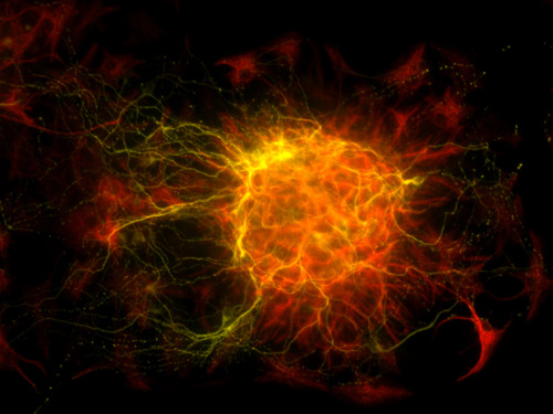
The Birth of Brain Cells
This might look like a distant web of galaxies captured by a powerful telescope, but it’s actually a microscopic image of a newborn nerve cell. The human brain contains more cells than there are stars in our galaxy, and the most important cells are neurons, which are nerve cells responsible for transmitting and processing electro-chemical signals at up to 320 km/h. This chemical signalling occurs through synapses—specialised connections with other cells, like wires in a computer. Each cell can receive input from thousands of others, so a typical neuron can have up to ten thousand synapses—i.e., can communicate with up to ten thousand other neurons, muscle cells, and glands. Estimates suggest that adult humans have approximately 100 billion neurons in their brain, but unlike most cells, neurons don’t undergo cell division, so if they’re damaged they don’t grow back—except, apparently, in the hippocampus (associated with memory) and the olfactory bulb (associated with sense of smell). The process by which this occurs is unclear, and this image was taken during a project to determine how neurons are born—it actually depicts newborn nerve cells in an adult mouse’s brain.
(Image Credit: Dana Bradford)


-
 bbowend0 liked this · 2 months ago
 -
 th3-0bjectivist liked this · 2 months ago
 -
 unforgettable-sensations liked this · 3 months ago
 -
 jamesmassino liked this · 3 months ago
 -
 dreamydespair liked this · 3 months ago
 -
 nudeartpluspoetry liked this · 3 months ago
 -
 hiromusicarts-blog liked this · 3 months ago
 -
 invincibleworld liked this · 3 months ago
 -
 bobcronkphotography liked this · 3 months ago
 -
 rodolfo9999 liked this · 3 months ago
 -
 paddy0121 liked this · 3 months ago
 -
 izmirliahmetkaya liked this · 3 months ago
 -
invincibleworld reblogged this · 3 months ago
-
 beforeyearning liked this · 3 months ago
 -
 injunjoeisticklish liked this · 4 months ago
 -
 tiramisu-muse liked this · 4 months ago
 -
 autonomy1 liked this · 4 months ago
 -
 stewacai liked this · 4 months ago
 -
 cuntsukuroi liked this · 4 months ago
 -
 inkndecho liked this · 4 months ago
 -
 kandybarre reblogged this · 4 months ago
 -
 juan-francisco-palencia liked this · 5 months ago
 -
 ziobbo liked this · 5 months ago
 -
 ninomeira liked this · 5 months ago
 -
 drmh2040 liked this · 6 months ago
 -
 24-04-toro liked this · 6 months ago
 -
 dinosaurwithablog liked this · 6 months ago
 -
 alex--max liked this · 6 months ago
 -
 meklarian liked this · 6 months ago
 -
 the-eternal-moonshine liked this · 6 months ago
 -
invincibleworld reblogged this · 6 months ago

Science nerd 🧪 | History buff 📜 | Dog & cat person 🐾always curious!
68 posts