Panic! At The Lab
panic! at the lab
ft hit songs such as
i write scribbles not lab reports
nine in the afternoon (nine in the evening? morning?) (oh it’s a 12 hr time point)
mad as grad students
high hopes (dissertation version)
More Posts from Thejoyofscience and Others

This bad little boy is super rare to find!!! This is a mitotically active cell present in peripheral blood circulation from a dog - only the second one I have ever seen!!
As a game, in the diagnostic lab we yell KABLAM! anytime we see a mitotic figure. The first one to say kablam wins :-P We should so turn it into a drinking game…

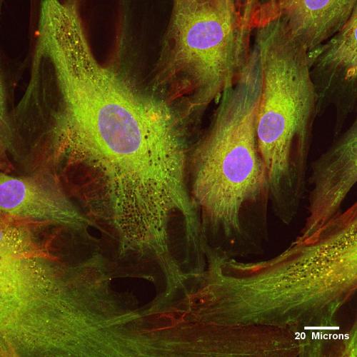
The central nervous system (CNS) in most vertebrates forms initially as a flat sheet of cells, which subsequently rolls up and fuses shut to form the hollow neural tube, which is the precursor to the CNS. The enriched apical actin in the closing neural tube (shown in green in the image) is central to cell shape changes that contribute to the rolling up process.
Image: Color micrograph showing a cross-sectional (transverse) view of the closing neural tube in a Xenopus embryo. Actin is shown in green.

Behold the Gastric Rainbow. Sounds gross, but it’s actually beautiful. This cross-section of a mouse intestine is labeled with a spectrum of fluorescent molecules. From the green and magenta digestive enzyme-producing cells to the red mucus-secreting cells, this is one of the most dynamic areas in the mammalian body: Each cell is replaced by another every 3-5 days.
(via The Scientist Magazine)

Thumbs Up For Science
The cover of the legendary journal Nature from February 1879, featuring this thumb microscope, yours for the low, low price of three pounds.
(via Ptak Science Books, which you should really check out)
For decades, scientists have been capitalizing off discoveries made from Henrietta Lacks’ family’s cells. That may change.


Ms. Holly Aaron, Dr. Karen Dehnert, Dr. Scott Laughlin, and Dr. Carolyn Bertozzi
University of California, Berkeley, California, USA Specimen: Fucosylated glycans in a zebrafish embryo. Technique: Confocal microscopy


“our work should equip the next generation of women to outdo us in every field this is the legacy we’ll leave.”
- rupi kaur
Hitchhiking bacteria might help their host navigate via magnetic fields
Deep in the mud of the Mediterranean Sea, scientists have caught microscopic protists dancing to a strange beat—the beat of Earth’s magnetic fields. Now, a new study reveals how these tiny clusters of cells orient themselves along those fields: by letting magneto-sensing bacteria hitch a ride on their outer membranes.
Researchers used microscopes to examine protist-packed sediment taken from the bottom of the Mediterranean Sea near Carry-le-Rouet, France. When they placed a magnet with its north pole facing a water droplet from the sediment, the hundreds of protists inside immediately began to swim toward the droplet’s edge. When the researchers reversed the magnet so its south pole was facing the droplet, the protists fled in the other direction (above).
Steering Stem Cells with Magnets
Magnets could be a tool for directing stem cells’ healing powers to treat conditions such as heart disease or vascular disease.
By feeding stem cells tiny particles made of iron oxide, scientists at Emory and Georgia Tech can use magnets to attract the cells to a particular location in the body after intravenous injection.
The results are published online in the journal Small and will appear in an upcoming issue.
Human Adipose Derived Mesenchymal Stem Cells
More…
-
 nuttyhologrammilkshake liked this · 4 years ago
 -
 reacom-draws reblogged this · 5 years ago
 -
 fictionvssleep reblogged this · 5 years ago
 -
 omganiyak liked this · 5 years ago
 -
 nagati liked this · 6 years ago
 -
 piromomo-blog liked this · 6 years ago
 -
 poolofpisces reblogged this · 6 years ago
 -
poolofpisces liked this · 6 years ago
-
 mb121314 reblogged this · 6 years ago
 -
mb121314 liked this · 6 years ago
-
 mythgirl07 reblogged this · 6 years ago
 -
 wot-gr liked this · 6 years ago
 -
 tagitables liked this · 6 years ago
 -
 ley-med liked this · 6 years ago
 -
 gottastaystudious liked this · 6 years ago
 -
 radpiewhispers reblogged this · 6 years ago
 -
 docresa reblogged this · 6 years ago
 -
 grqficfeelings liked this · 6 years ago
 -
 nicktheking9999-blog liked this · 6 years ago
 -
 queenofthebench liked this · 6 years ago
 -
 dhuntwork liked this · 6 years ago
 -
 thejoyofscience reblogged this · 6 years ago
 -
 tomatostaches-turtlebabies liked this · 6 years ago
 -
ggyinvxtu-blog liked this · 6 years ago
-
 johnnys-so liked this · 6 years ago
 -
 be-the-change-jess reblogged this · 6 years ago
 -
 skiesfullofsong liked this · 6 years ago
 -
 com-itten liked this · 6 years ago
 -
 astudyinphd reblogged this · 6 years ago
 -
 kiseki94 reblogged this · 6 years ago
 -
kiseki94 liked this · 6 years ago
-
 roseh23 liked this · 6 years ago
 -
 sophia42888 liked this · 6 years ago
 -
 animepanda51 reblogged this · 6 years ago
 -
animepanda51 liked this · 6 years ago
-
ladadidooo2 liked this · 6 years ago
-
 melinda-reads liked this · 6 years ago
 -
 realkermit liked this · 6 years ago
 -
 avichyssoise reblogged this · 6 years ago
 -
 epalzeorhynchos-frenatum reblogged this · 6 years ago
 -
 akanbaetsunemori liked this · 6 years ago
An assortment of scientific things from the wonderful world of biology
77 posts