Connective Tissue

Connective tissue
Anne Weston, London Research Institute, Cancer Research UK
This false-coloured scanning electron micrograph shows connective tissue removed from a human knee during arthroscopic surgery. Individual fibres of collagen can be distinguished and have been highlighted by the creator using a variety of colours. The horizontal field width of the image is 16 microns.
More Posts from Thejoyofscience and Others


Aspirate of a mammary mass from a 8 year-old, female-intact, Yorkshire Terrier. The patient was found wondering in a field by a good Samaritan-turned-owner over the 4th of July weekend. Although she was acting normally, the owner brought the little dog in for a ‘look over.’ On physical examination a 2cm mass was felt in the left mammary chain. No obvious spay scar was present.
*
On cytology there were copious clusters of epithelial cells. These epithelial cells (top picture) were very non-descript, making for large, jumbled piles of cells. Notice how you cannot see any well-defined cell borders between them?? Just a ton of nuclei (and nucleoli) blending in together! That’s a sign of cell craziness! Many clusters were surrounded by this gorgeous, pink-magenta material. Likely secretory product or matrix.
*
Cytologic diagnosis: Mammary tumor! Although the cells look quite malignant on cytology, many studies have shown you cannot reliably determine malignancy with cytology alone. Thus, you NEED a biopsy to determine if a mammary tumor is malignant or benign in a dog. And flip a coin on that - about 50% are malignant and 50% are benign. Intact female dogs have an almost 35% lifetime chance of developing one of these beasts!

This bad little boy is super rare to find!!! This is a mitotically active cell present in peripheral blood circulation from a dog - only the second one I have ever seen!!
As a game, in the diagnostic lab we yell KABLAM! anytime we see a mitotic figure. The first one to say kablam wins :-P We should so turn it into a drinking game…

Thumbs Up For Science
The cover of the legendary journal Nature from February 1879, featuring this thumb microscope, yours for the low, low price of three pounds.
(via Ptak Science Books, which you should really check out)


Amazing Annual Monarch Butterfly Migrations
Monarch butterflies in other countries also migrate with the season, but it’s those in North America that travel the greatest distance. Each year, there are two major Monarch Butterfly migrations in North America. Those monarchs that live east of the Rocky Mountains fly down to Mexico, while the more western population stops in California. Monarchs do not like the cold, and as soon as things start to get a little chilly up north, they take off south (and west) for warmer climates.
The largest group travels over 1,250 miles from the Rocky Mountains to spend the winter in Michoacán, Mexico. The government of Mexico has managed to almost stamp out logging in the monarch’s wintering areas, a practice which once threatened the migrating insects. Working with environmental organizations and individuals, they have been encouraging communities to start eco-tourism enterprises by planting trees for the butterflies to nest. The monarch is a butterfly ruled by the sun. When the autumn sun reaches fifty two degrees above the horizon, the monarch reproduction cycle shuts down, and their great migration begins. When they begin their flight down to Mexico, they have never been there, yet every generation is able to find the exact same spot year after year where their previous ancestors spent the winter.
The second group travels from Ontario, Canado to spend their winters in Santa Cruz, California. You may wonder why the monarchs don’t simply stay and enjoy the warmer weather there year round. That’s because they need the milkweed plants on which their larvae feed, and those are more plentiful up north. So as soon as the weather starts to warm up, that’s where they return every year. Interestingly, not every generation of monarchs migrate. Some simply remain in their breeding ground. Those that do migrate are born at the end of summer or early autumn. Because of their trip to warmer climes, this special generation will outlive several younger generations that stay put. It will then be the migratory monarchs’ great grandchildren that follow the beat of their forebears’ wings.
in Ontario, Canada, in their summer home. It’s thought that the distinctive bright coloring of the monarchs acts as a warning to predators to stay away. Monarch butterflies are also poisonous and will make any animal that tries to eat them sick – hopefully sick enough not to try snacking on them a second time! The poison comes from the milkweed that they eat while they are caterpillars. This doesn’t always work, however. Certain bird species, for example, have learned that some parts of the butterflies are not as toxic, while other predators are resistant or immune to the poison altogether.
source


Journey to the Microcosmos- The Complicated Legacy of Lynn Margulis
Images originally captured by Jam’s Germs
This is probably silly, but would you mind breaking down the genders of bees in the hive and some of the roles the various genders take on? I've heard mixed things about what gender drones are, males dying immediately after sex with the queen (?), and I'm wondering how and when the hive produces another queen! Thank you so much, your patience and knowledge are hugely appreciated. Also, are there any cool insects that can change sex like frogs and fish occasionally can?
There is no such thing as gender in animals besides humans and gender roles aren’t based on anything other then misogyny and they don’t exist within animals.
What you’re specifically asking about is called castes, and your typical eusocial species of bee such as a honeybee have three castes; workers, drones and a queen.

A queen’s function within the hive is to lay eggs, while a drones only purpose is to mate. The workers are tasked with every other role within the colony including cleaning, disposing of the dead or diseased, nursing, guarding and protecting the hive, foraging for pollen and nectar, and so on.
The queen lays two types of eggs; haploid (half the amount of chromosomes which is 16 in bees) and diploid (both sets of 32 chromosomes) eggs. The haploid eggs become drones meaning that they have no fathers, this is a type of parthenogenesis called arrhenotoky.
The normal diploid eggs (chromosomes sets from the queen + one of the drones that mated with her) can become either workers or a new queen. Every single diploid egg has the potential to become a queen. It is diet in the larval stage that determines if the egg will be a queen or a worker.
The workers will create a new queen themselves by building a specific brood comb called a queen cup, once the old queen has laid an egg in them, the workers will begin feeding them entirely on royal jelly: a secretion from the hypopharynx of nurse worker bees while larvae that will become workers are fed some royal jelly but mostly a mixture of pollen and nectar called bee bread. Because the royal jelly is so protein-rich it provides the future queen larvae with the energy to develop into a sexually mature females.

Once the queen emerges from her cell fully-mature she will seek out other recently hatched virgin queens and unemerged queens and killed them as they are rivals: there can only be one queen after all. When the virgin queen is the sole survivor she’ll then leave the hive for the first (and often the last and only time) for her mating flight. Bees mate on the wing, so swarms of drones will chase after her competing for the chance to mate with the single queen, these are called drone comets.
If a drone is lucky enough to mate with the queen, he’ll insert his penis into her before it is torn off (and often left inside the queen). This causes the drones to fall to the ground and die shortly after. The queen will mate with multiple drones sporing their sperm inside an organ called the spermatheca in which she uses to fertilise and lay her eggs.
Here’s a queen with the penis of a drone still plugged inside her, otherwise known as the mating sign.

Not all drones get this chance to mate, and often die outside the hive because they’re evicted for reasons like lack of resources, the hive not being healthy / strong, or it’s the end of the bee season going into winter. Drones are relatively expendable within the hive and are useless at feeding themselves so they die pretty quickly.

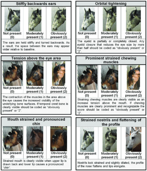

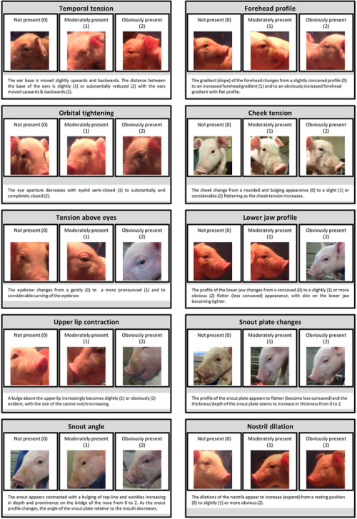


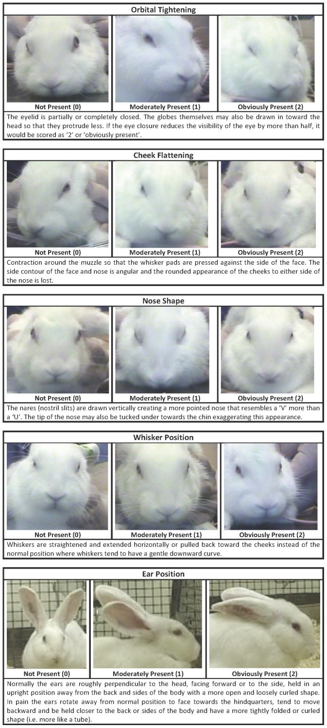

Recognising silent acute pain in animals - assorted species grimace scales:
Development of the Horse Grimace Scale (HGS) as a Pain Assessment Tool in Horses Undergoing Routine Castration
The composition and initial evaluation of a grimace scale in ferrets after surgical implantation of a telemetry probe
The Assessment of Facial Expressions in Piglets Undergoing Tail Docking and Castration: Toward the Development of the Piglet Grimace Scale
The Sheep Grimace Scale as an indicator of post-operative distress and pain in laboratory sheep and the Coding and quantification of a facial expression for pain in lambs
Mouse - How to be a pain management advocate for exotic and zoo animals (full text available - includes additional species)
The Rat Grimace Scale: A partially automated method for quantifying pain in the laboratory rat via facial expressions
Evaluation of EMLA Cream for Preventing Pain during Tattooing of Rabbits: Changes in Physiological, Behavioural and Facial Expression Responses
Pain evaluation in dairy cattle
Pain is subtle - we cannot depend on vocalisations or extreme abnormal behaviour to determine if an animal is on pain - animals can cover up pain while going about their daily life. Grimace scales have been found to be reliable indicators of pain (full text available)
Unfortunately, I could not find a clear visual grimace scale for dogs, cats or birds :(
Which is a shame, because perhaps I could have recognised my own dog’s discomfort for the acute pain it was sooner:

(left: dog in pain. See eyes, tension, cheeks, whiskers, ears compared to the multiple species grimace charts above. right: tired but not in pain dog)
Perhaps my new books that arrived today might have some on dogs at least. There’s this visual blog post of a stressed dog at the vet - stress in the absence of a trigger looks very much like pain.
Here is a small comparative cats, with the link going into more detail. Not a scale but better than nothing:


Bonus round - you can get free A3 posters on recognising pain for Rabbits, Mice and Rats from the National Centre for the Replacement, Refinement and Reduction of Animals in Research. My rabbit specialist vet has the rabbit one!


This is Phthirus pubis, a human louse, commonly known as “crabs" — it is known to infect pubic hair but can infect the EYELASHES.
I came across this while making a high yield vector borne disease lecture for an upcoming talk. On the clinical pathology boards, they apparently really like to show “gross" gross photographs of bugs and ask what is it or what disease does it cause. I have a fascination with tick born disease, so it felt natural to expand and learn some more entomology. My attending was looking through my lecture and said I needed to add an example of a louse and a flea, since people commonly mix them up. Well, I naturally started to google… and ended up grossing myself AND my attending out.
A job well done. Another excellent day on clinical microbiology.
PS - up until a year ago, I thought that “crabs" were actually miniature crabs. What? I wasn’t that far off! Both are apart of the phylum arthropoda!
New project!
I received a new research project in my lab today on concurrent ehrlichia infections in dogs! All the happy feels!
-
 roussii liked this · 9 years ago
 -
 ah-thenah reblogged this · 9 years ago
 -
 katiekateeeee08 reblogged this · 9 years ago
 -
 homotology reblogged this · 9 years ago
 -
 quiltsandspg liked this · 9 years ago
 -
 sylfir reblogged this · 10 years ago
 -
 tommes14 reblogged this · 11 years ago
 -
tommes14 liked this · 11 years ago
-
 femmeviva liked this · 11 years ago
 -
 growingseason reblogged this · 11 years ago
 -
 luxarchitek reblogged this · 11 years ago
 -
 yuruyurau reblogged this · 11 years ago
 -
 afilamelcolmillo reblogged this · 11 years ago
 -
 existentialismandmakeup liked this · 11 years ago
 -
 wetwareontologies liked this · 11 years ago
 -
 silicongene liked this · 11 years ago
 -
 dermoosealini reblogged this · 11 years ago
 -
 itsgoodtoseayou-blog liked this · 11 years ago
 -
 qiujingtian liked this · 11 years ago
 -
 robertflemingjones reblogged this · 11 years ago
 -
 ixchebelyax-blog-blog reblogged this · 11 years ago
 -
 nap-toon liked this · 11 years ago
 -
 taoofpaschx liked this · 11 years ago
 -
 baldelfmage-archive reblogged this · 11 years ago
 -
 brainsandbodies reblogged this · 11 years ago
 -
brainsandbodies liked this · 11 years ago
-
 onceuponavictor reblogged this · 11 years ago
 -
 drekkin reblogged this · 11 years ago
 -
 polymath4ever reblogged this · 11 years ago
 -
 hotdogcephalopod reblogged this · 11 years ago
 -
 molecularlifesciences reblogged this · 11 years ago
 -
 lineadecuatro reblogged this · 11 years ago
An assortment of scientific things from the wonderful world of biology
77 posts