Journey To The Microcosmos- The Complicated Legacy Of Lynn Margulis


Journey to the Microcosmos- The Complicated Legacy of Lynn Margulis
Images originally captured by Jam’s Germs
More Posts from Thejoyofscience and Others


Usual interstitial pneumonia
It has the loose fibroblastic balls (pink balls) next to relatively normal lung parenchyma (thin septae) and a bronchiole (the blueish wrinkly thing)
(Via pulmonary pathology)
Medical lung is like… impossible. COP. DAD. DIP. NSIP. BOOP. POOP. SCHMOOP. No idea. It all looks the same to me.


Aspirate of a mammary mass from a 8 year-old, female-intact, Yorkshire Terrier. The patient was found wondering in a field by a good Samaritan-turned-owner over the 4th of July weekend. Although she was acting normally, the owner brought the little dog in for a ‘look over.’ On physical examination a 2cm mass was felt in the left mammary chain. No obvious spay scar was present.
*
On cytology there were copious clusters of epithelial cells. These epithelial cells (top picture) were very non-descript, making for large, jumbled piles of cells. Notice how you cannot see any well-defined cell borders between them?? Just a ton of nuclei (and nucleoli) blending in together! That’s a sign of cell craziness! Many clusters were surrounded by this gorgeous, pink-magenta material. Likely secretory product or matrix.
*
Cytologic diagnosis: Mammary tumor! Although the cells look quite malignant on cytology, many studies have shown you cannot reliably determine malignancy with cytology alone. Thus, you NEED a biopsy to determine if a mammary tumor is malignant or benign in a dog. And flip a coin on that - about 50% are malignant and 50% are benign. Intact female dogs have an almost 35% lifetime chance of developing one of these beasts!

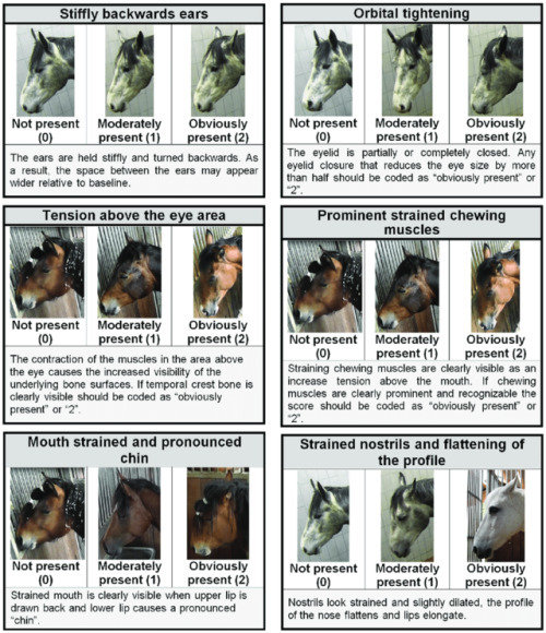

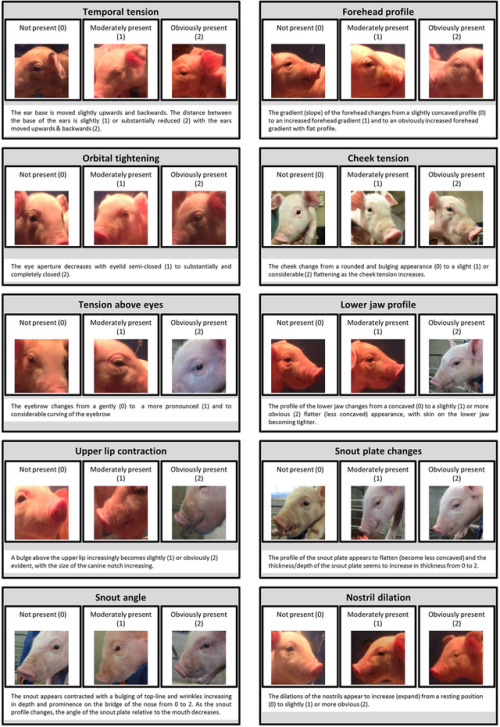


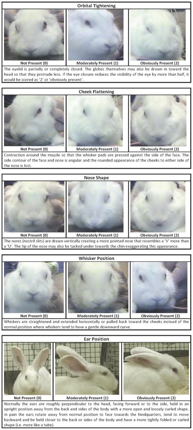

Recognising silent acute pain in animals - assorted species grimace scales:
Development of the Horse Grimace Scale (HGS) as a Pain Assessment Tool in Horses Undergoing Routine Castration
The composition and initial evaluation of a grimace scale in ferrets after surgical implantation of a telemetry probe
The Assessment of Facial Expressions in Piglets Undergoing Tail Docking and Castration: Toward the Development of the Piglet Grimace Scale
The Sheep Grimace Scale as an indicator of post-operative distress and pain in laboratory sheep and the Coding and quantification of a facial expression for pain in lambs
Mouse - How to be a pain management advocate for exotic and zoo animals (full text available - includes additional species)
The Rat Grimace Scale: A partially automated method for quantifying pain in the laboratory rat via facial expressions
Evaluation of EMLA Cream for Preventing Pain during Tattooing of Rabbits: Changes in Physiological, Behavioural and Facial Expression Responses
Pain evaluation in dairy cattle
Pain is subtle - we cannot depend on vocalisations or extreme abnormal behaviour to determine if an animal is on pain - animals can cover up pain while going about their daily life. Grimace scales have been found to be reliable indicators of pain (full text available)
Unfortunately, I could not find a clear visual grimace scale for dogs, cats or birds :(
Which is a shame, because perhaps I could have recognised my own dog’s discomfort for the acute pain it was sooner:

(left: dog in pain. See eyes, tension, cheeks, whiskers, ears compared to the multiple species grimace charts above. right: tired but not in pain dog)
Perhaps my new books that arrived today might have some on dogs at least. There’s this visual blog post of a stressed dog at the vet - stress in the absence of a trigger looks very much like pain.
Here is a small comparative cats, with the link going into more detail. Not a scale but better than nothing:


Bonus round - you can get free A3 posters on recognising pain for Rabbits, Mice and Rats from the National Centre for the Replacement, Refinement and Reduction of Animals in Research. My rabbit specialist vet has the rabbit one!


Right lateral thoracic wall mass from a 10 year-old, female-spayed, Golden Retriever. Approximately six months ago the owner noticed a small, firm swelling on the patient’s right chest. No doubt it grew with time, as it finally got large enough for the owner to become truly concerned! The patient is clinically healthy otherwise.
*
The most prominent feature on the cytology were these gorgeous globules of magenta, streaming material! In fact, the entire slide was imbued with this color when you looked at it without a microscope. The substance is most likely matrix produced by cancer cells. Matrix of this color is commonly observed in chrondrosarcomas. Occasional malignant spindle-shaped cells were found imbedded in the matrix.
*
Diagnosis: Chondrosarcoma. Well , that’s the most likely diagnosis given the location and appearance of the neoplastic cells and matrix! Other possibilities include a myxosarcoma (which produces a mucous like material!) or osteosarcoma (malignant primary bone tumor). Chondrosarcomas are often regionally invasive but rarely metastasize. If the owner is on board with a chest wall resection, the patient will have a good prognosis :-)

Kim Weaver (b. 1964) is an astrophysics professor and astronomer. She is an expert in x-ray astronomy and has worked for NASA’s Goddard Space Flight Center.
She obtained her PhD in astronomy in 1993 from the University of Maryland. After that, she was a research scientist at Penn State and John Hopkins University. Her honours include the Presidential Early Career Award and the NASA Peer Award.
underappreciated form of humor: using incorrect long forms of proper names i.e. Craigory, Bobert, Barold, etc.
I just read that bees don't have lungs how do their respiratory system works??
Correct. Insects do not have lungs they breathe through spiracles (tiny openings) that can open and close, as well as having filters to keep dust and other external contaminants out.

The spiracles run across their abdomen connecting to tracheae which is where oxygen exchange takes place (In mammals this happens in the blood), that then connect to tracheole. The air sacs expand and collapse so force the air in the spiracles and into the trachea and almost function in that they can reserve air in them so insects can conserve water.

Honey bees have 10 pairs of spiracles, 3 pairs on the thorax and 7 pairs on the abdomen. Bees can use and accelerate the passage of air in their bodies via their air sacs, contacting them and increasing their respiration rate.
This adaptive function of their respiratory system actually helps them to fly and the first few spiracles are used for air to exit while the others for air entering, they also have valves to prevent backflow. This also allows the bee to cool down or heat up if it needs to, which is why you’ll see bees abdomens wiggling while they’re resting. They’re forcing air in and out of their bodies in order to rapidly cool down or heat up, which is important for flight.


The central nervous system (CNS) in most vertebrates forms initially as a flat sheet of cells, which subsequently rolls up and fuses shut to form the hollow neural tube, which is the precursor to the CNS. The enriched apical actin in the closing neural tube (shown in green in the image) is central to cell shape changes that contribute to the rolling up process.
Image: Color micrograph showing a cross-sectional (transverse) view of the closing neural tube in a Xenopus embryo. Actin is shown in green.


the himalayan monal is a large member of the pheasant family found in parts of asia. while during the breeding season they mainly stay in pairs, in winter they form small communities and roost together. they feed on grasses, insects, seeds and berries. they are known for their vivid iridescent plumage, particularly colorful for a pheasant. x
-
 legendoftherisingtide reblogged this · 2 years ago
 -
legendoftherisingtide liked this · 2 years ago
-
 sophies-junkyard liked this · 2 years ago
 -
 cicadaland reblogged this · 2 years ago
 -
 pheygele reblogged this · 2 years ago
 -
pheygele liked this · 2 years ago
-
 m-e-w-666 reblogged this · 2 years ago
 -
m-e-w-666 liked this · 2 years ago
-
 seven-oculi liked this · 3 years ago
 -
 mvampirequeen reblogged this · 3 years ago
 -
 algaeequeen reblogged this · 3 years ago
 -
algaeequeen liked this · 3 years ago
-
 takineko reblogged this · 4 years ago
 -
 gothopera reblogged this · 4 years ago
 -
gothopera liked this · 4 years ago
-
 hereforthefanart liked this · 4 years ago
 -
 scoopertainment-talks liked this · 4 years ago
 -
 camisadeforcapreta liked this · 4 years ago
 -
 fdivitor reblogged this · 4 years ago
 -
 thewindwaker reblogged this · 4 years ago
 -
 sartres-angst reblogged this · 4 years ago
 -
sartres-angst liked this · 4 years ago
-
 lavendercokezero liked this · 4 years ago
 -
 romissilva reblogged this · 4 years ago
 -
 sergeanthunderfists liked this · 4 years ago
 -
romissilva liked this · 4 years ago
-
 albedobeheading reblogged this · 4 years ago
 -
albedobeheading liked this · 4 years ago
-
 vampireteefs reblogged this · 4 years ago
 -
 chupgopigon liked this · 4 years ago
 -
 captntrashcan13 liked this · 4 years ago
 -
 blueskyswoons liked this · 4 years ago
 -
 getsherlock221b liked this · 4 years ago
 -
 bluemongerpaperrebel liked this · 4 years ago
 -
 mightyalex liked this · 4 years ago
 -
 dw001 liked this · 4 years ago
 -
 leoforpeace-blog liked this · 4 years ago
 -
 ser-fae liked this · 4 years ago
 -
 iamanemotionaltimebomb reblogged this · 4 years ago
 -
iamanemotionaltimebomb liked this · 4 years ago
-
 fangtasium liked this · 4 years ago
 -
 wxrldwithoutru1es reblogged this · 4 years ago
An assortment of scientific things from the wonderful world of biology
77 posts